- Surgery
- Pets
VSC Update: Lincoln | Spontaneous Pneumothorax
May 22, 2024
One recent Saturday morning, the owners of Lincoln, an 8-year-old active German Short-haired Pointer, noticed that he was not acting himself. The owners took him to a local emergency clinic where a spontaneous pneumothorax was diagnosed. A spontaneous pneumothorax is a serious condition in which air enters the space in the chest called the pleural space, between the surface of the lungs and the inside of the rib cage. This causes the lungs to collapse.
Lincoln’s owners met Dr. Rose and a CT scan of the chest was scheduled for that same day. A CT scan (or CAT scan) allows for visualization of the lungs and internal organs.


The CT revealed that Lincoln had a condition called a pulmonary bulla. A bulla is a thin-walled, air-filled pocket in the lungs that can burst, allowing air to enter into the chest cavity. Lincoln had over 3 liters of air in his chest collapsing his lungs! The treatment for the condition requires the surgeon to evaluate each of the lung lobes to find the hole in the surface and remove that segment of the lungs. Typically, this procedure involves open chest surgery, and the recovery is very long (like heart bypass surgery for a human).
But the doctors at VSC had another option for Lincoln, minimally invasive thoracic surgery! Using a camera and a special anesthesia device, the lungs could be selectively deflated and checked through small incisions in the chest. VSC is the only clinic on the west coast of Florida that routinely performs this type of procedure!
Until surgery could be performed Lincoln had chest drains to prevent accumulation of air. He returned to VSC for surgery with Dr. Rose and Dr. Peress. During the procedure the hole in the lung was identified (see video below). During the delivery of breaths under anesthesia the leak of air can be found by looking for a stream of bubbles leaking from the submerged lung. The lung was successfully removed with a special stapling device that sealed the end of the damaged lung. He recovered from surgery with the chest tubes but they were removed the next day as he showed no recurrence of the pneumothorax. Because of the minimally invasive surgery he quickly recovered from surgery with minimal pain.

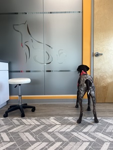
Lincoln recently had his post-surgery follow up and he is doing very well. His owners also say that he is doing well at home and is back to playing with his two fur-siblings. The tissue was sent to the lab for pathology. Lucky for Lincoln the lung tissues appeared healthy in all areas but the bulla. All of us at VSC are so happy that Lincoln is doing well and wish him all the best in the future! We are truly so honored that we had the opportunity to care for such an amazing dog!